Research Peptides — 6-Panel Analytical Testing, Every Lot
We don’t ask you to
take our word for it.
Every lot of our research peptides undergoes six independent analytical tests before it ships. Every result goes into the public COA library before the product leaves our facility — not after you request it.
Switching vendors? See what we carry →
| Test | Result |
|---|---|
| 01 Identification | 1419.5 Da |
| 02 Purification | 99.1% |
| 03 Conformity | Conforms |
| 04 Sterility | Absent |
| 05 Quantification | Confirmed |
| 06 Endotoxin | 0.02 EU/mL |
Issued Mar 09, 2026
Culver City, CA
US Made Research Peptides
Synthesized, lyophilized, and finished in U.S. facilities. Every domestic lot passes the same six-panel analytical testing as the rest of the catalog, with its certificate of analysis published.
USACJC-1295 (no DAC)+Ipamorelin (5+5)
Snapshot CJC-1295 No DAC (GHRH analog) and ipamorelin (GHS-R1a agonist) stimulate endogenous GH through complementary cAMP and Ca²⁺ signaling pathways. Clinical and mechanistic studies of GHRH+GHS-R1a agonists co-administration demonstrate amplified pulsatile GH release compared to either pathway alone, supporting dual-axis activation of the somatotroph system. What Is CJC-1295 Without DAC? CJC-1295 without DAC is a synthetic analog of growth hormone–releasing hormone (GHRH amino acid 1–29 or Mod GHF1-29 ). This CJC-1295 is designed to stimulate endogenous growth hormone (GH) secretion through activation of the GHRH receptor (GHRH-R) on anterior pituitary somatotrophs. Without the Drug Affinity Complex (DAC), it does not bind albumin, resulting in a shorter half-life and more physiologic, pulse-like GH stimulation. Mechanistically, it activates the adenylyl cyclase–cAMP–protein kinase A (PKA) signaling cascade, promoting GH release and downstream increases in circulating insulin-like growth factor-1 (IGF-1) [1]. Because it works upstream at the hypothalamic–pituitary axis, CJC-1295 without DAC preserves endogenous inhibitory feedback regulation via somatostatin and IGF-1. What Is Ipamorelin? Ipamorelin is a selective growth hormone secretagogue (GHS) that binds to the ghrelin receptor (GHS-R1a) on anterior pituitary somatotrophs [2]. It stimulates endogenous GH release primarily through activation of the phospholipase C (PLC)–IP3–calcium signaling pathway, increasing intracellular calcium and promoting pulsatile GH secretion [3]. Unlike earlier GHS compounds and ghrelin itself, ipamorelin is relatively selective for GH release, with minimal stimulation of ACTH, cortisol, or hunger compared to less selective secretagogues [2]. By acting through a pathway distinct from GHRH analogs, ipamorelin is frequently studied in combination paradigms evaluating complementary stimulation of the somatotroph axis. Synergy CJC-1295 without DAC and ipamorelin stimulate endogenous growth hormone (GH) release through distinct but convergent regulatory pathways within the HPA axis. Their combined use is based on dual activation of GHRH and ghrelin receptor systems via: GHRH receptor > adenylyl cyclase–cAMP–PKA (CJC-1295) GHS-R1a > phospholipase C (PLC)–IP3–Ca²⁺(Ipamorelin) Because these pathways operate independently, their combined activation should increase both the magnitude and efficiency of GH pulsatility. Although published studies typically evaluate GHRH combined with GHRP compounds (such as GHRP-6 or ghrelin) rather than ipamorelin specifically, the mechanistic framework likely applies to ipamorelin due to its selective GHS-R1a agonism. Cell-based studies demonstrate that co-activation of GHRH and GHS receptors can produce approximately twofold greater cAMP signaling compared to GHRH alone, suggesting receptor-level cross-talk and amplification of somatotroph responsiveness [4]. A clinical study evaluated whether ghrelin, the endogenous ligand for the GHS receptor, interacts synergistically with growth hormone–releasing hormone (GHRH) to stimulate GH secretion [5]. 8 male adults were administered ghrelin (0.08, 0.2, and 1.0 μg/kg) intravenously alone or combined with 1.0 μg/kg GHRH. Results showed that combined administration with GHRH: Produced significantly greater GH responses than either peptide alone (p < 0.05)[a] GH response exceeded the sum of the individual responses, demonstrating true supra-additive synergy (p < 0.050 No synergistic interaction with ACTH or prolactin secretion This study demonstrates that co-administration of ghrelin and GHRH produces true synergistic GH release in humans, exceeding additive stimulation from either agent alone. The findings support the concept that dual activation of the GHRH receptor and GHS receptor enhances pituitary somatotroph responsiveness. In some metabolic conditions (e.g., obesity-associated blunting of GHRH response), GHS agonists partially restored GH responsiveness [6]. Ipamorelin Pairing Ipamorelin is highlighted in research contexts due to its relative selectivity for GH release, with minimal stimulation of ACTH and cortisol compared to earlier GHRP compounds. This selective profile may allow more targeted evaluation of somatotroph activation without broader pituitary axis activation. When paired with a short-acting GHRH analog such as CJC-1295 without DAC, the goal is typically to: Preserve physiologic pulsatility Enhance GH pulse amplitude Maintain endogenous feedback regulation The absence of the DAC component in CJC-1295 results in a shorter half-life, aligning more closely with natural episodic GH dynamics rather than prolonged elevation. Dose and Ratio Considerations Balanced ratios such as 2 mg + 2 mg or 5 mg + 5 mg can be conceptually described as targeting simultaneous engagement of: The GHRH-R/cAMP axis (transcriptional and secretory priming) The GHS-R1a/Ca²⁺ axis (secretory amplification) Proportional dosing may theoretically promote coordinated receptor activation. However, precise optimization of dose ratios has not been definitively established in controlled combination trials and remains an empirical parameter in research settings. References: 1 Sackmann-Sala, L., Ding, J., Frohman, L. A. and Kopchick, J. J. (2009) Activation of the GH/IGF-1 axis by CJC-1295, a long-acting GHRH analog, results in serum protein profile changes in normal adult subjects. Growth Horm. IGF Res., Elsevier BV 19, 471–477 2 Raun, K., Hansen, B. S., Johansen, N. L., Thøgersen, H., Madsen, K., Ankersen, M., et al. (1998) Ipamorelin, the first selective growth hormone secretagogue. Eur. J. Endocrinol., Oxford University Press (OUP) 139, 552–561 3 Mear, Y., Enjalbert, A. and Thirion, S. (2013) GHS-R1a constitutive activity and its physiological relevance. Front. Neurosci., Frontiers Media SA 7, 87 4 Cunha, S. R. and Mayo, K. E. (2002) Ghrelin and growth hormone (GH) secretagogues potentiate GH-releasing hormone (GHRH)-induced cyclic adenosine 3’,5'-monophosphate production in cells expressing transfected GHRH and GH secretagogue receptors. Endocrinology, The Endocrine Society 143, 4570–4582 5 Hataya, Y., Akamizu, T., Takaya, K., Kanamoto, N., Ariyasu, H., Saijo, M., et al. (2001) A low dose of ghrelin stimulates growth hormone (GH) release synergistically with GH-releasing hormone in humans. J. Clin. Endocrinol. Metab., The Endocrine Society 86, 4552 6 Popovic, V., Damjanovic, S., Micic, D., Djurovic, M., Dieguez, C. and Casanueva, F. F. (1995) Blocked growth hormone-releasing peptide (GHRP-6)-induced GH secretion and absence of the synergic action of GHRP-6 plus GH-releasing hormone in patients with hypothalamopituitary disconnection: evidence that GHRP-6 main action is exerted at the hypothalamic level. J. Clin. Endocrinol. Metab., The Endocrine Society 80, 942–947 [a]As currently written, only this first bullet point grammatically fits the intro sentence. "Combined administration" is functioning as the subject, and "produced" is the verb. The other two don't begin with similar verbs (and have different structures). Needs to be harmonized, and there are multiple ways you could do that, making sure that each bullet matches the intro. Also: There is an issue in that the bullets indicate 3 variations: ghrelin alone, GHRH alone, and both combined. But the description above only says "ghrelin alone or combined with GHRH." That's only 2 variations. There is no GHRH alone mentioned above.
USAMOTS-c
What is MOTS-c peptide? MOTS-c is a mitochondrial-derived and exercise-induced peptide whose levels decrease with age [1]. It improves insulin sensitivity, glucose metabolism, and metabolic homeostasis [2]. Animal studies suggest that, by activating AMPK, it can mitigate obesity resulting from high-fat diets, aging, and menopause [3, 2]. It also regulates age-related inflammation and various aspects of age-related physical decline, such as bone and muscle losses [1]. MOTS-c peptide benefits and side effects Metabolic and cardiovascular health In pancreatic cells, MOTS-c lowers insulin secretion and increases glucagon production [4]. In mice with aberrant lipid metabolism, MOTS-c treatment significantly reduced lipid buildup in liver cells [5]. In rodents, high-fat diet-induced obesity can promote insulin resistance and fat accumulation that triggers chronic inflammation. In mice, MOTS-c administration protects against both age-related and high-fat diet-induced insulin resistance, and diet-induced obesity [6]. Hormonal changes during pregnancy can affect insulin sensitivity and cause high blood sugar, leading to gestational diabetes. In pregnant women and mouse models of gestational diabetes, MOTS-c normalized blood sugar, and enhanced insulin sensitivity and glucose tolerance. The MOTS-c treatment lowered both the birth weight-associated complications and mortality of offspring caused by gestational diabetes [7]. Low estrogen during menopause can cause weight gain and fat redistribution in favor of white fats, which increases insulin resistance and the risk of metabolic disorders. In contrast, the more mitochondria-dense brown fats burn more calories, and improve glucose and lipid metabolisms. In ovarectomized mice, MOTS-c increases brown fat activation, and reduces fat accumulation and inflammation in white adipose tissue, which contributes to the lower level of fats in serum and liver [3]. In heart failure, fluid buildup and increased pressure in the lungs can cause lung injury. MOTS-c reduced heart dysfunction and remodeling caused by heart failure and lowered inflammation while boosting antioxidant activity in the hearts of sick mice [8]. It can also prolong injured heart and lung cells’ life cycle [9, 10]. Muscle building and bone health Age-related sarcopenia can reduce overall healthspan and independence, while increasing various metabolic health risks. MOTS-c suppresses myostatin and lipid infiltration that contributes to muscle atrophy, even in immobilized animals [11, 12]. Older adults with higher circulating MOTS-c have better muscle performance and lean mass, while those with lower levels experienced more sarcopenia [13]. In a mouse model of aging, MOTS-c can significantly increase physical performance by activating genes related to skeletal muscle metabolism and myoblast adaptation to metabolic stress [1]. In an in vitro model of muscle development, MOTS-c peptide helps muscle cells to form properly, affecting their integral parts such as myotubes. It supports muscle cell formation at the expense of lipid accumulation while protecting muscle cells from the breakdown effects of inflammatory cytokine IL-6 [14]. Age-related bone loss often parallels muscle loss, and various anti-sacropenic stimuli also protect bone health. Importantly, metabolic dysfunction and inflammation tend to accelerate bone loss. MOTS-c mitigates age-related bone loss by stimulating osteoblasts and suppressing osteoclasts by modulating AMPK and inflammatory responses [15]. In a mouse bone damage model, MOTS-c treatment reduced bone loss and inflammation and prevented the formation of osteoclasts [16]. Anti-inflammatory and antioxidant benefits Sepsis is a potentially lethal condition characterized by systemic overactive immune reactions towards an infection or noninfectious agents. MOTS-c greatly improves survival and lowers bacterial counts in MRSA-infected experimental mice. It also reduces levels of pro-inflammatory cytokines like TNF-α, IL-6, and IL-1β, while increasing the anti-inflammatory cytokine IL-10 [17]. Type 1 diabetes patients have lower endogenous MOTS-c levels than healthy controls. In a mouse model of type 1 diabetes, exogenous MOTS-c prevents pancreatic β cell destruction by shifting CD4+ T cells towards a less self-destructive phenotype, suggesting that MOTS-c may be beneficial as an autoimmune treatment [18]. Age-related inflammation and declining antioxidant capacity are key drivers of aging and related diseases. Older adults aged 70–81 years have 20% less MOTS-c than younger adults 18–30 years old [19]. In a mouse model of type 2 diabetes, MOTS-c administration increased antioxidant enzymes like SOD and CAT, protecting myocardial cells from oxidative stress [20]. Neuropathic pain Unlike acute pain, neuropathic pain is pain caused by nervous system dysfunction related to nerve damages, rather than by injury or inflammation. MOTS-c’s antinociceptive effects pertain to its ability to restore mitochondrial health, and inhibit microglial and pain signals in the spinal cord [21]. Furthermore, compared to morphine, MOTS-c has far fewer side effects such as gastrointestinal transit inhibition and motor incoordination [22].
USAEpitalon
What is epitalon peptide? Epitalon, also known as Epithalone or Epithalon, is a synthetic tetrapeptide (Ala–Glu–Asp–Gly) derived from a naturally occurring pineal gland extract (epithalamin) [1]. Structurally, epitalon mimics endogenous peptides that influence the activity of telomerase, the enzyme responsible for maintaining telomere length. Epitalon may also modulate oxidative stress regulation, circadian rhythm stabilization, and neuroendocrine function. Epitalon peptide benefits Anti-aging and longevity One of the most widely studied aspects of epitalon is its potential influence on cellular aging through multiple biochemical pathways. A cell study investigated whether Epithalon can influence two processes central to dementia, cholinesterase activity and the formation of the soluble form of amyloid precursor protein (sAPP) in human neuroblastoma (SH-SY5Y) cells [2]. Results showed that Epithalon: Reduced excessive cholinesterase enzymatic activity. Increased sAPP formation, which is protective against Alzheimer’s-type pathology. These effects suggest that epitalon may delay or prevent mechanisms underlying conditions like Alzheimer’s disease. Similar anti-aging effects are also seen in other tissues, such as in the prevention of age-based pigmentary retinal dystrophy in genetically predisposed rats [3]. Antioxidant Oxidative stress is a central contributor to cellular aging and carcinogenesis, as unmanaged oxidative species can damage lipids, proteins, and DNA. Epitalon can mitigate such damages by enhancing endogenous antioxidant defenses. In a fruit fly study, synthetic Epithalon’s antioxidant activities were compared with the crude pineal extract, epithalamin [4]. Epitalon was added to larval nutrient medium at 0.00001 wt%, while epithalamin was added at concentrations 1000-fold higher. Epitalon addition resulted in: 20% Increased catalase activity (p<0.05) 20-50% decreased CHP content (marker of lipid peroxidation) (p<0.05) ~1000-fold higher biological activity than epithalamin Anticarcinogenic A study evaluated the effect of epitalon on tumor development and oncogene expression in 80 transgenic HER-2/neu mice, a model predisposed to breast cancer and accelerated aging [5]. Mice were either given saline as negative control, Vilon as a positive control, or Epithalon (1 µg, subcutaneous) for 5 consecutive days monthly. Results showed that epitalon: Delayed first tumor appearance by 38 days compared to Vilon, and by 20 days compared to saline. Reduced recurring tumor incidence: 28% remained tumor-free vs.18% (saline). Lowered tumor multiplicity: only 56% had ≥2 tumors vs. 75% (saline). Reduced maximum tumor diameter by 33% (p<0.05). 3.7-fold lower HER-2/neu mRNA expression compared to saline (p<0.001), whereas Vilon produced a 1.97-fold lower HER-2/neu expression. Telomere protection A defining feature of epitalon is its reported influence on telomerase activity. Telomerase is the ribonucleoprotein enzyme responsible for elongating telomeric DNA, maintaining chromosomal stability. In most somatic cells, telomerase activity is repressed, which leads to progressive telomere shortening during replication. When telomeres become critically short, cells enter senescence, a hallmark of aging [6]. An in vitro study evaluated whether epitalon can activate telomerase and elongate telomeres in human somatic cells [7]. Human fetal fibroblasts were exposed to epitalon at varying concentrations, resulting in: Increased telomerase Telomere elongation Extended proliferative capacity beyond the normal Hayflick limit (40-60 replications before senescence and death) Circadian rhythm Epitalon has been linked to pineal gland regulation through melatonin, a hormone that regulates sleep–wake cycles and seasonal biological rhythms. Melatonin naturally declines with age. An animal study evaluated whether epitalon can restore melatonin secretion and normalize circadian rhythms of cortisol production in aged female rhesus monkeys [8]. The experimental group received 10 µg of epitalon intramuscularly, once daily for 10 days, while the control group received saline. epitalon administration resulted in: A three-fold increase in evening melatonin secretion (48 vs. 15 pg/ml, p < 0.001) Restoration of normal day-night variations of circadian cortisol rhythm Effects selective for aged animals only Metabolic regulation Age-related changes in mitochondria, nutrient sensing, and hormone signaling may underpin metabolic changes that result in insulin resistance and other related diseases. Epitalon works partly by addressing these mechanisms, according to a study in rhesus monkeys [9]. Seven young (6–8 years) and seven old (20–27 years) monkeys were administered epitalon intramuscularly (10 µg/day for 10 days). After administration, older monkeys had: Decreased basal glucose concentration Improved glucose clearance rate (p<0.01) Restored early-phase insulin secretion (320% vs. 198% control, p<0.05) Improved late-phase insulin dynamics Glucose tolerance improvements persisted for 1–2 months post-treatment, even after epitalon withdrawal.
Compound Catalog
Lyophilized powder. Research-grade compounds with full analytical testing on every lot.
Standard lab runs use 3–4 peptides per protocol. Orders over $150 ship free — most multi-peptide orders qualify.

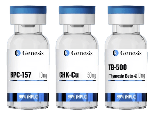
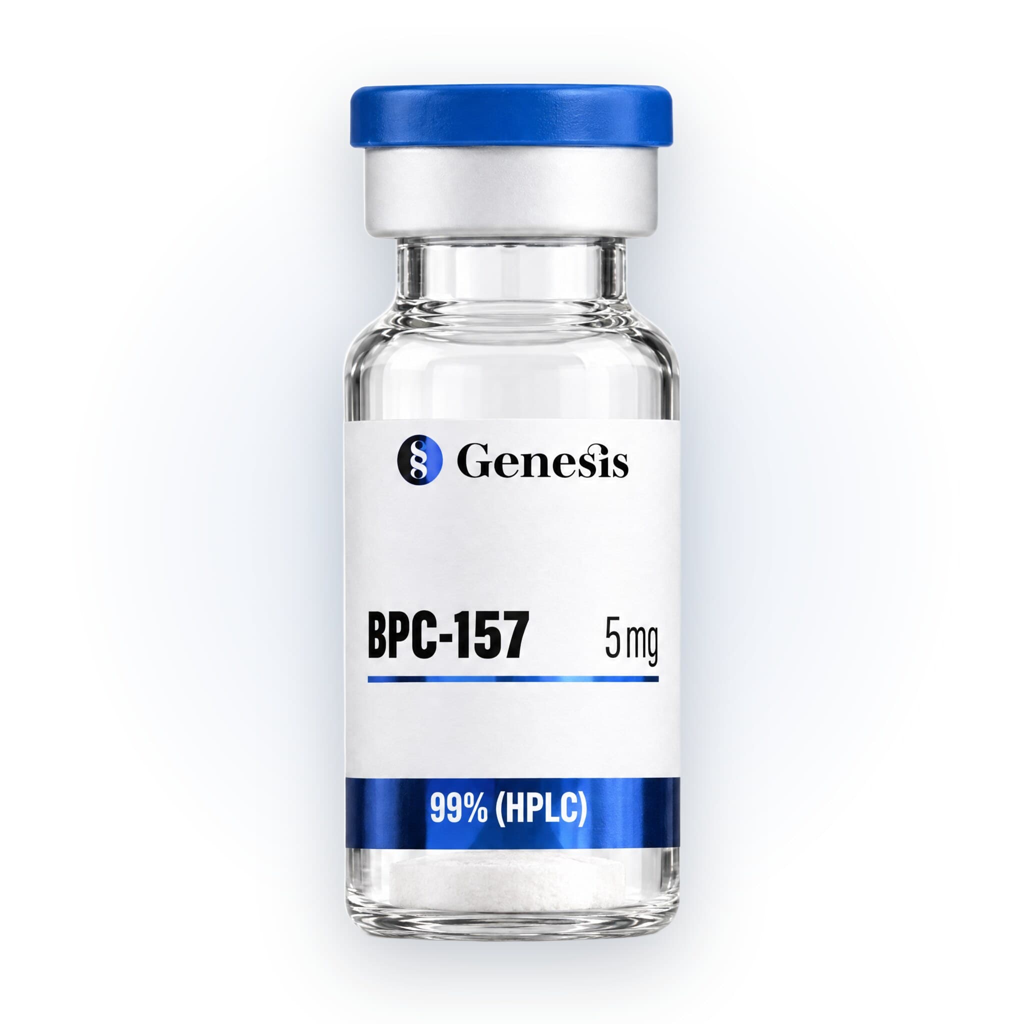
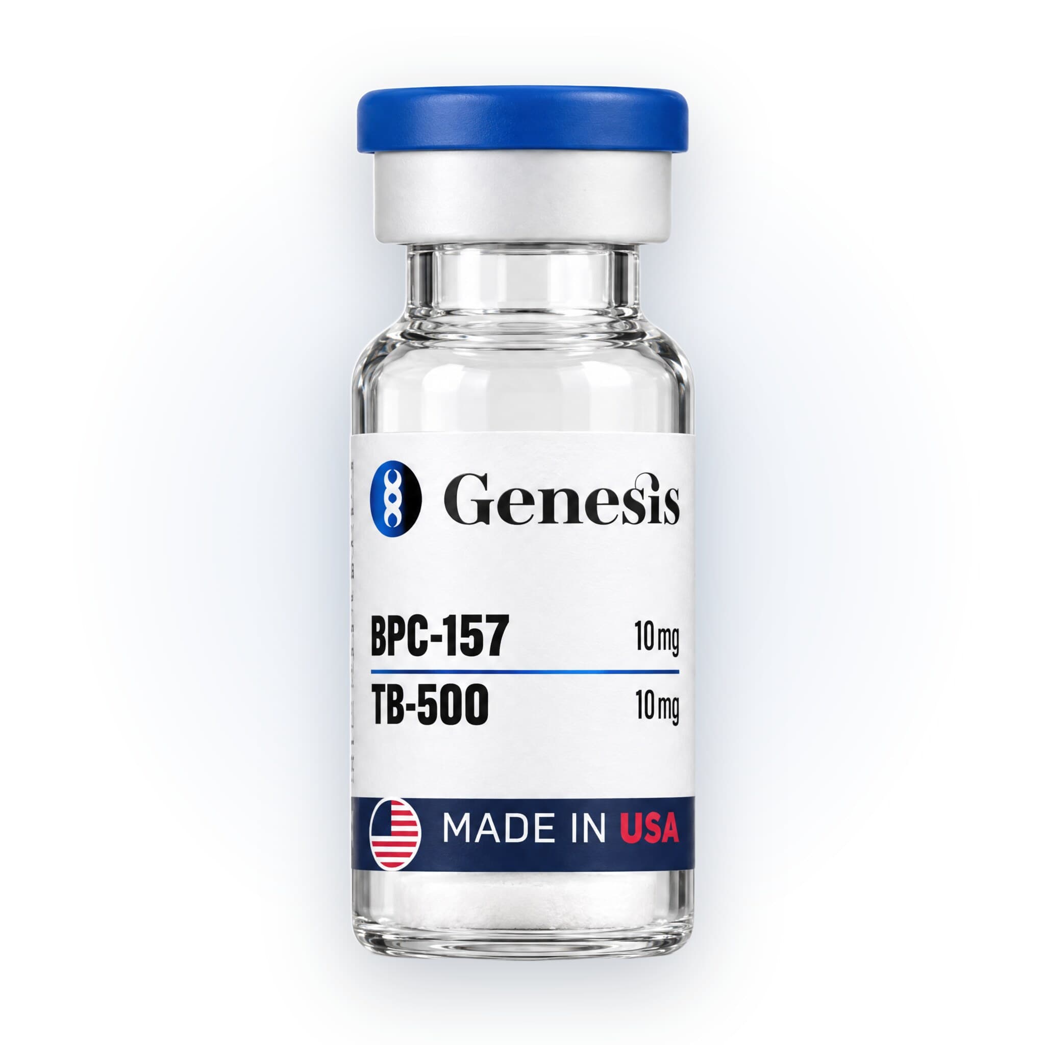
BPC-157
What is BPC-157? BPC-157 is a synthetic pentadecapeptide composed of 15 amino acids, with the sequence Gly-Glu-Pro-Pro-Pro-Gly-Lys-Pro-Ala-Asp-Asp-Ala-Gly-Leu-Val. It is a portion of a protein that occurs naturally in human gastric juice, commonly referred to as Body Protection Compound (BPC). Native BPC helps maintain gastrointestinal integrity under normal physiological conditions. BPC-157 is stable and soluble in water. As a synthetic peptide, BPC-157 is not produced endogenously in this exact form but is designed to replicate a biologically active fragment of the parent protein found in gastric secretions [1]. The peptide has been the subject of extensive preclinical research, particularly in the context of cellular proliferation, angiogenesis, and tissue repair mechanisms. BPC-157 has been studied in animal and in vitro models as a tool for investigating pathways associated with: Gastrointestinal homeostasis Vascular modulation Cytoprotection Inflammation modulation All research involving BPC-157 remains within the domain of experimental studies, and it is not approved for human therapeutic use. What does BPC-157 do? The research Inflammation and pain modulation BPC-157 has been extensively studied in preclinical models for its modulatory effects on inflammation, particularly in relation to tissue injury and repair processes [1]. In various rodent studies, administration of BPC-157 reduced markers of inflammation in models of gastrointestinal, musculoskeletal, and neural injury. By modulating inflammation, rat studies suggest that various BPC variants modulate pain, while BPC-157 predominantly reduces acute pain in incisional and formalin-induced pain [2]. In a rat model of allodynia, it also reduced pain by protecting nerve integrity from capsaicin [3]. BPC may also modulate the nitric oxide (NO) system. It counteracts both excessive and deficient NO activity, supporting endothelial integrity and attenuating leukocyte infiltration in inflamed tissues. This dual regulation may contribute to its observed ability to balance pro-inflammatory and anti-inflammatory signaling cascades [4]. In experimental colitis models, for example, BPC-157 administration was associated with: Reduced mucosal damage Decreased myeloperoxidase activity (a marker of neutrophil infiltration) Normalization of cytokine profiles BPC-157 also promotes angiogenesis (blood vessel growth) and stabilizes vascular function at sites of injury. This angiogenic support not only facilitates tissue repair but may also limit secondary inflammation resulting from ischemia and oxidative stress [5]. Research in tendon and ligament injury models similarly highlights reductions in edema and inflammatory cell presence following BPC-157 exposure. These findings are derived from animal studies, and while promising, they await confirmation in human clinical research to fully understand its therapeutic relevance. Tissue healing and stem cells BPC-157 can facilitate regeneration across various tissue types, including: Tendon Muscle Ligament Bone Nerve A key feature identified in these models is BPC-157’s capacity to modulate cellular and immune environments in ways that promote structural integrity and restoration of injured sites. In rat models of tendon fibroblasts, BPC-157 significantly upregulated growth hormone receptor expression, and enhanced the responsiveness of these cells to endogenous growth hormone [6]. This interaction promotes cell proliferation and tissue regeneration, increasing expression of proliferation markers: Proliferating cell nuclear antigen (PCNA) JAK2 signaling pathway These results suggest a supportive role in tendon repair processes at the molecular level, enhancing the body's natural regenerative mechanisms. BPC-157 may influence stem cell activity indirectly by optimizing the local microenvironment of injured tissues. While direct stimulation of stem cell differentiation by BPC-157 has not been definitively proven, its actions on surrounding tissues, blood vessels, and extracellular matrix components are believed to indirectly enhance stem cell-mediated repair processes. Gut health BPC-157 has been extensively studied in preclinical models for its protective and regenerative effects on the gastrointestinal (GI) tract [7]. Research in rat models of ileoileal anastomosis healing, for instance, shows that BPC-157: Modulates local immune responses Enhances granulation tissue formation Increases collagen and reticulin deposition Promotes re-epithelialization Supports the regeneration of muscular tissue strands at anastomotic sites. BPC-157 reduced adhesion formation and necrosis, while accelerating the resolution of edema and inflammatory infiltrates in treated animals compared to controls. Additional experimental models of intestinal injury, including those involving perforations, fistulas, or induced colitis, report that BPC-157 administration supported gut integrity by: Promoting angiogenesis Mitigating tissue necrosis Stabilizing microvascular structures The peptide’s influence on nitric oxide pathways and its modulation of endothelial function are proposed mechanisms underlying these benefits. While clinical trials in humans remain limited, the consistent findings across animal models offer a compelling basis for future investigation into its application in gut health contexts. BPC-157 in bees Bees are crucial pollinators whose populations are in significant declines. Research suggests that BPC-157 may help to improve bee health and survival. In bees, BPC-157 improves colony strength and enhances certain aspects of immune responses [8]. Supplementation of bee diets with the peptide reduced the infection load of the Nosema ceranae, a one-celled fungal parasite. BPC-157 also attenuates gut damage from N. ceranae infections [8]. Overall, BPC-157 may be beneficial in beekeeping.

Thymosin Beta-4
What is TB500 (Thymosin Beta 4)? TB500 peptide is a shorter, bioactive fragment of thymosin beta-4, designed to focus on thymosin beta-4’s most therapeutically relevant region, the 7-amino acid sequence LKKTETQ responsible for actin binding and tissue regeneration [1]. Though structurally simpler than the full-length thymosin beta-4, TB500 peptide retains potent biological activity [5]. The number 500 in TB500 is added as a commercial name, without biological or scientific significance. Key biochemical features include: Actin-binding domain [6]: Essential for cytoskeletal regulation, enabling cell migration and tissue remodelling. Essential amino acid residues [6]: Support actin polymerization and cellular mobility. No post-transitional modifications [6]: As a synthetic peptide, TB500 lacks glycosylation or phosphorylation, ensuring structural stability. What does TB500 do? TB500 exerts multi-system effects, supporting wound healing, reducing inflammation, promoting cell regeneration, and enhancing immune defenses [1]. Tissue repair and regeneration TB500 accelerates tissue repair by binding actin, a key structural protein in cells. This interaction stimulates stem cell recruitment and differentiation at injury sites, migration of skin cells to close wounds faster, and formation of new blood vessels (angiogenesis) to improve oxygen and nutrient delivery [1]. It also enhances collagen alignment and increases laminin-5, both essential for strong and well-structured tissues [7]. Simultaneously, it reduces the number of scar-forming cells, minimizing fibrotic tissue formation [8]. Animal studies have confirmed TB500 peptide’s ability to reduce tissue damage, speed up recovery, and promote healing even in challenging conditions [9]. Human trials suggest that topical formulations are safe and effective in wound repair. Emerging data also support the potential role of TB500 in neurological and cardiac tissue regeneration, aiding recovery after events like stroke or heart attack [9]. Anti-inflammatory and immunomodulatory effects Following tissue injury, high levels of inflammation can damage tissues and lead to permanent scarring. TB500 peptide mitigates this response, lowering the levels of inflammatory cells and the chemical signals they release [1]. This has downstream impacts of reducing tissue swelling, protecting healthy tissue, and creating an environment supportive of proper healing with less scar tissue formation [1]. A key anti-inflammatory mechanism involves the NF-kB signalling pathway, which controls the expression of many pro-inflammatory genes. TB500 inhibits NF-kB activation, prevents p65 subunit phosphorylation, and blocks nuclear translocation of NF-kB [10]. These actions have been demonstrated in corneal, cardiac, and liver tissues. The NF-kB inhibition contributes to reduced inflammation and improved healing responses in these tissues [10]. TB500 peptide modulates the toll-like receptor-4 (TLR-4) pathway, which is central to innate immune responses [11]. Through upregulation of microRNA-146a, TB500 can suppress this pathway, promoting anti-inflammatory effects [11]. For this reason, TB500 may indirectly support the repair of gut barriers by improving the proliferation and migration of cells, and supporting tissue healing processes. Furthermore, a peptide fragment within TB500, Ac-SDKP, has been shown to reduce fibrosis (e.g., heart scarring after myocardial infarction), likely through similar anti-inflammatory and anti-proliferative mechanisms [6]. Antibacterial and antiviral effects TB500 strengthens antimicrobial defenses by increasing the expression of antimicrobial peptides (AMPs) such as keratin 6A, CAMP, beta-defensins (BD2, BD3), and S100A8 [12]. These peptides help prevent bacterial adherence and enhance immune clearance of pathogens [12]. TB500 also boosts TLR4 expression, enhancing the recognition of bacterial invaders like LPS-producing pathogens [12]. When combined with antibiotics, TB500 enhances the activity of 12-LOX and 15-LOX enzymes, which promote resolution of inflammation and tissue restoration [12]. This synergy highlights TB500’s potential as an adjunct to antimicrobial therapies—supporting not only microbial defense but also repair of infected tissues.

GHK-Cu
What is GHK-Cu? Native GHK-Cu peptide is released from the extracellular matrix, cleaved from its parent protein, SPARC (secreted protein, acidic, and rich in cysteine), during extracellular matrix remodelling. Studies find it has vital roles in copper transport, tissue repair, inflammation modulation, and gene regulation [1]. A tripeptide, GHK-Cu is composed of glycine (Gly), histidine (His), and lysine (Lys) [1]. This amino acid complex binds tightly to copper, forming a stable coordination complex [2]. This binding capacity is what gives GHK-Cu its unique biological signalling and regenerative properties [2]. After performing its functions, GHK-Cu is degraded by serum peptidases. Studies show that circulating levels decrease with age, averaging 200 ng/mL at age twenty and declining to 80 ng/mL by age sixty, potentially impacting regenerative capacity [1]. Copper(II) is a redox-active metal required as a cofactor for many enzymes, including cytochrome C oxidase, lysyl oxidase, and superoxide dismutase [2]. GHK binds copper in a bioavailable, non-toxic form, supports shuttling it into cells, and maintains intracellular copper homeostasis [2]. GHK-Cu shows abilities to up- or downregulate over 4,000 genes. These pathways are many, and include those involved in tissue regeneration and repair, anti-inflammatory signalling, antioxidant defenses, and cell growth and differentiation [2]. While scientists are still elucidating some of GHK-Cu peptide’s mechanisms, current research suggests that they include augmenting transcription factors, epigenetic modification (particularly histone and chromatin) and oxidative stress signalling [2]. What does GHK-Cu do? The research Research is still emerging on the wide applications of GHK-Cu peptide; however, our current understanding of its actions point towards applications particularly in skin and hair, and regenerative processes. GHK-Cu and skin Research suggests that GHK-Cu peptide may have roles in reprogramming older tissues to act more youthful [2]. When skin gets injured, GHK-Cu is naturally released, acting as an emergency signal to activate skin healing processes [3]. A study using a test tube wound model found that GHK-Cu increases the production of collagen (which gives skin structure), elastin (which keeps skin elastic), and decorin and glycosaminoglycans (key molecules that hydrate and organize skin tissue) [4]. GHK-Cu also supports the balance of enzymes that break down skin proteins, MMPs, and their inhibitors, TIMPs [5],[6]. This prevents the buildup of damaged proteins, and overactive breakdown, which can lead to thinning and sagging skin [2]. One cell-based study found that skin cells exposed to GHK-Cu in combination with red LED light had 12.5x greater cell survival, 230% increase in fibroblast growth factor, and 70% higher collagen production [7]. At the stem cell level, GHK-Cu improved the health and shape of basal cells in the epidermis, increasing markers of stemness, which may help skin regenerate better with age [8]. GHK-Cu and hair GHK-Cu peptides show benefits for supporting hair growth, by [9]: Improving blood flow to hair follicles Preventing hair shedding and premature hair loss Stimulating the growth of new hair follicles Studies find that GHK-Cu can stimulate fibroblasts, a particular type of skin cell, to produce VEGF (vascular endothelial growth factor), which helps support the growth of new blood vessels surrounding the hair follicle [9]. More blood vessels allow for greater nutrient and oxygen delivery to the area, thus supporting stronger, faster growing hair [9]. It also reduces the production of TGF-beta, a chemical that signals to hair follicles to stop growing and enter the shedding phase of hair growth [9]. With less TGF-beta, hair remains in the growth phase for longer. By encouraging dermal papilla cells to multiply and protecting them from apoptosis, GHK-Cu supports the development and growth of new hair, by keeping follicles healthy [9]. Other regenerative processes While more research is needed, our understanding of GHK-Cu points towards potential applications of this peptide in wound healing and repair processes, promotion of antioxidant defenses, angiogenesis, gene modulation, and more [2].
Free standard shipping on orders over $150 · Same-day dispatch before noon PST, Mon–Sat · For research use only

Why we run six tests when most vendors run one.
Analytical methods validated against pharmaceutical industry acceptance criteria.
Electrospray ionization mass spectrometry. Mass confirmed to ±0.1 Da of theoretical. Multiply charged ion envelope deconvoluted for intact mass.
Molecular identity. Confirms the synthesized peptide matches the intended sequence by mass. Spectrum published in COA.
A peptide can show 99% chromatographic purity and still be the wrong compound. Mass spec confirms molecular identity independently.
Reversed-phase HPLC with UV detection at 220 nm. Purity by area normalization. Minimum acceptance: ≥98.0%.
Total peptide purity. Identifies co-eluting impurities, incomplete couplings, and degradation products. Chromatogram published with every COA.
Confirms purity but not identity. A mis-synthesized peptide with the wrong sequence can read ≥99% pure. That is why identification by mass spec is required as a second check.
Visual inspection of the lyophilized cake and container closure, with label and lot reconciliation against the batch record.
That what ships matches what was released — correct compound, correct fill, intact vial, and a lot number that traces to the batch record.
Analytical data describes the material that was assayed. Conformity confirms the vial in the box is that same material.
Microbial enumeration and screening for specified organisms per USP <61> / <62>. Membrane filtration with plate count.
Viable bacterial and fungal load. Distinct from endotoxin — a lot can be endotoxin-clean and still carry live organisms introduced after synthesis, during lyophilization or fill.
Requires incubated culture over several days, which delays lot release. Invisible to HPLC, mass spectrometry, and the LAL assay alike.
Acid hydrolysis (6N HCl, 110°C, 24 hrs) followed by chromatographic quantification of amino acid ratios. For peptides >15 residues.
Net peptide content and amino acid composition. Confirms correct residues in expected proportions.
Without it, dosing is based on gross weight including TFA counterions and moisture. Quantification provides the actual peptide fraction for accurate dosing.
Limulus amebocyte lysate (LAL) assay per USP <85>. Kinetic turbidimetric method. Acceptance: ≤ 0.05 EU/mL.
Bacterial endotoxin (lipopolysaccharide). Pyrogenic and can confound inflammatory endpoints in vivo, regardless of peptide purity.
Requires dedicated instrumentation and validated depyrogenation. Invisible to both HPLC and mass spectrometry.
Tests 4–6 require dedicated instrumentation and culture facilities beyond standard HPLC/MS and are not part of most research peptide vendors’ standard release panel. All analytical data, including raw spectra and measured values, is published in the COA for each lot.
Certificates of Analysis — published before shipment, searchable by lot number, downloadable without account creation.
Every lot documented before it ships — not after you request it. Every result in the public library.
| LOT | PRODUCT | HPLC PURITY | DATE |
|---|
SHOWING 0 OF — LOTS
Lot numbers on your COA match the lot numbers in this library. If a lot number is not listed, contact us before using the compound.
Search All COAs by Lot NumberResearch Institutions and Lab Teams
University labs, CROs, and biotech teams order through us with documentation formatted for institutional procurement review.
- — Volume pricing starting at 100+ units
- — Chemists available for institutional support
- — COAs formatted for records management and IRB documentation
Genesis Peptides operates from Culver City, California. Every certificate of analysis is downloadable without an account, searchable by lot number, and includes verification data from our third-party testing laboratory. We have maintained continuous operations with a clean regulatory record.